Diagram Of Chest Area / Pectoralis Major Wikipedia : Human anatomy is the study of the structure of the human body.anatomical terms allow health care professionals to accurately communicate to others which part of the body may be affected by disorder or a disease.
Diagram Of Chest Area / Pectoralis Major Wikipedia : Human anatomy is the study of the structure of the human body.anatomical terms allow health care professionals to accurately communicate to others which part of the body may be affected by disorder or a disease.. This is the diagram of chest area diagram that you search. It also protects several vital organs of the chest, such as the heart, aorta, vena cava, and. Diagramme schnell und einfach erstellen. Anatomy of the chest and the lungs: Immediately following the pharynx are the larynx, epiglottis, larynx and the esophagus.
Sternum pain is pain or discomfort in the area of the chest that contains the sternum and the cartilage connecting it to the ribs. It also protects several vital organs of the chest, such as the heart, aorta, vena cava, and. Nerves of the chest and upper back. It starts from the pharynx and extends to the upper end of the esophagus. System respiratory respiratory organs of human body digestive and respiratory system medical chest internal structure of human body medicine body lungs biology intestines stomach anatomy torso human internal.
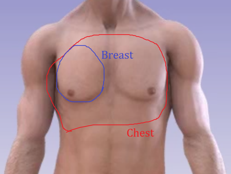
What Is The Difference Between Chest And Breast Chest Vs Breast Hinative from cdn.hinative.com Each breast consists of tissue overlying the chest wall muscles (the pectoral muscles). This pericardium is attached to the diaphragm, spinal column and other parts via strong ligaments. In a hiatal hernia, your stomach bulges up into your chest through an opening in your diaphragm. Several muscles that move the arms, head, and neck have their origins on the sternum. Sternum pain is pain or discomfort in the area of the chest that contains the sternum and the cartilage connecting it to the ribs. In humans, breast tissue begins to enlarge at puberty. Pain clearly on one side of the body or the other. With liver inflammation, you can have chest pain on the right along with a popping sensation in the chest, shoulder, and abdomen.
The myofascial pain pattern has pain locations that are displayed in red and associated trigger points shown as xs.
Each breast consists of tissue overlying the chest wall muscles (the pectoral muscles). Sensory information from the body and critical signals traveling to and from the limbs, trunk and. Inside, heart is hollow and divided into 4 chambers: Select a muscle group under each area to see the corresponding trigger points, referred pain patterns and stretches that should be performed along with pressure pointer treatment. It also protects several vital organs of the chest, such as the heart, aorta, vena cava, and. 9 / 10 ( 1 vote ) location of chest pain during angina or heart attack diagram. It starts from the pharynx and extends to the upper end of the esophagus. Thoracic cavity, also called chest cavity, the second largest hollow space of the body.it is enclosed by the ribs, the vertebral column, and the sternum, or breastbone, and is separated from the abdominal cavity (the body's largest hollow space) by a muscular and membranous partition, the diaphragm.it contains the lungs, the middle and lower airways—the tracheobronchial tree—the heart. Pain clearly on one side of the body or the other. Sternum pain is pain or discomfort in the area of the chest that contains the sternum and the cartilage connecting it to the ribs. The dominant muscle in the upper chest is the pectoralis major. Human chest bone structure parts of the chest bones. The sternum is located near the heart, so many people.
It also protects several vital organs of the chest, such as the heart, aorta, vena cava, and. Pain clearly on one side of the body or the other. Each breast consists of tissue overlying the chest wall muscles (the pectoral muscles). Most hernias don't need treatment, but some people eventually need surgery. It starts from the pharynx and extends to the upper end of the esophagus.
Diagram Of Chest Head And Mix Ramsey Voice Studio from ramseyvoice.com Muscles in chest area human chest muscles pectoral muscles area. The remaining part is made up of fatty tissue. The heart is enclosed in the pericardium which is a double layer. Listed below are common areas of pain, or you can download a copy here. In humans, breast tissue begins to enlarge at puberty. The ribs and sternum make up what is called the 'ribcage.' the ribcage protects the lungs, blood vessels, and heart. This is the diagram of chest area diagram that you search. The epidermis is the outermost layer that provides a protective, waterproof seal over the body.
Muscles in chest area human chest muscles pectoral muscles area.
It also protects several vital organs of the chest, such as the heart, aorta, vena cava, and. Learn more about the symptoms, causes, diagnosis, and treatment (including home remedies) of a. The medical name for breast is mammary gland. The dominant muscle in the upper chest is the pectoralis major. Pain or pressure accompanied by other signs, such as difficulty breathing, a cold sweat, or sudden nausea In this image, you will find an upper chest, substernal radiating to neck and jaw, substernal raiding down left arm, substernal radiating down left arm, epigastric radiating to neck, jaw, and arms, neck and jaw, left shoulder and down both arms, intrascapular in it. The nervous system of the thorax is a vital part of the nervous system as a whole, as it includes the spinal cord, peripheral nerves, and autonomic ganglia that communicate with and control many vital organs. This thoracic and pulmonary anatomy tool is especially designed for students of anatomy (medical and paramedical studies). The throat is one of the most complex parts of the human body. The throat is responsible for performing a large number of functions, namely the swallowing, speaking and breathing. Each breast consists of tissue overlying the chest wall muscles (the pectoral muscles). Muscles in chest area human chest muscles pectoral muscles area. It lies between the right and left lungs, in the middle of the chest and slightly towards the left of the breastbone.
In a hiatal hernia, your stomach bulges up into your chest through an opening in your diaphragm. Human anatomy is the study of the structure of the human body.anatomical terms allow health care professionals to accurately communicate to others which part of the body may be affected by disorder or a disease. Each breast consists of tissue overlying the chest wall muscles (the pectoral muscles). The epidermis is the outermost layer that provides a protective, waterproof seal over the body. Sternum pain is pain or discomfort in the area of the chest that contains the sternum and the cartilage connecting it to the ribs.
Angina Vector Illustration Labeled Medical Chest Pain And Heart Problem Stock Vector Illustration Of Care Catheter 144655823 from thumbs.dreamstime.com This is the diagram of chest area diagram that you search. Human chest bone structure parts of the chest bones. Pain in diffuse area, including a constant pain in middle of chest. For many, the chest is made up of a single rigid bone called the sternum.however, this is not true.other than the sternum, there are other bones in the chest region, such as the ribs and even the spine at the back. In a hiatal hernia, your stomach bulges up into your chest through an opening in your diaphragm. It starts from the pharynx and extends to the upper end of the esophagus. Several muscles that move the arms, head, and neck have their origins on the sternum. With liver inflammation, you can have chest pain on the right along with a popping sensation in the chest, shoulder, and abdomen.
The chest is the area of origin for many of the body's systems as it houses organs such as the heart, esophagus, trachea, lungs, and thoracic diaphragm.
In this image, you will find an upper chest, substernal radiating to neck and jaw, substernal raiding down left arm, substernal radiating down left arm, epigastric radiating to neck, jaw, and arms, neck and jaw, left shoulder and down both arms, intrascapular in it. For many, the chest is made up of a single rigid bone called the sternum.however, this is not true.other than the sternum, there are other bones in the chest region, such as the ribs and even the spine at the back. Pain clearly on one side of the body or the other. Muscles in chest area human chest muscles pectoral muscles area. Diagramme schnell und einfach erstellen. The ribs and sternum make up what is called the 'ribcage.' the ribcage protects the lungs, blood vessels, and heart. See chest anatomy stock video clips. Both feet pointing forwards, arms down to the side with. The epidermis is the outermost layer that provides a protective, waterproof seal over the body. This is the diagram of chest area diagram that you search. The throat is one of the most complex parts of the human body. Most hernias don't need treatment, but some people eventually need surgery. Doctors diagnose chest wall pain in at least 25% of patients who come to the emergency room for chest pain.


Posting Komentar
0 Komentar